This article was originally posted by HDYONZ [http://www.hdyo.co.nz/research-1/2016/8/21/deep-inside-the-huntingtons-brain]
Movement issues are one of the most commonly known symptoms of Huntington’s disease. If a person is familiar with the condition, they will know about the excessive uncontrolled movements (chorea) and eventual immobility (akinesia) caused by the disease.
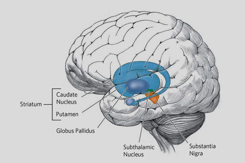
These symptoms are caused by the death of brain cells called medium spiny neurons. These cells connect two areas of the brain that are important for movement. The medium spiny neurons get their information from the striatum, which is a very important motor area, and these cells send the information to the globus pallidus. There are three sections of the globus pallidus: the internal section which sends movement information to other areas of the brain, the external section which regulates your movement system, and the ventral pallidum which controls movement in response to motivation and reward.
Researchers know that when the medium spiny neurons start to die, the globus pallidus as a whole begins to shrink, but they hadn’t figured out why it was shrinking. In this research, scientists were able to examine each part of the globus pallidus and determine which parts were being affected and how they were being affected.
In this study, researchers from The University of Auckland used brain tissue from the donated brains of people who had Huntington’s disease and compared it to tissue from the donated brains of people who had no known issues with their brains. These brains are located in the Centre for Brain Research, at The University of Auckland. By comparing the tissue from these two groups, the scientists were able to see what the cells and tissue of a healthy globus pallidus looked like and were able to compare that to what they saw when they examined the globus pallidus from the brains of the individuals with Huntington’s disease.
What they found was that the different parts of the globus pallidus were impacted differently. The external section and the ventral pallidum were the most severely affected. The globus pallidus externus showed a 54% reduction in total size, 60% loss of neurons, and 34% reduction in the size of the neurons which had survived. The ventral pallidum had a 31% reduction in total size, 48% loss of neurons, and a 64% reduction in the size of surviving neurons. In comparison, the globus pallidus internus only showed a reduction of 38% in its total size but no other real differences from the healthy brain tissue.
This proves that each area of the globus pallidus is affected differently by Huntington’s disease, and so each area will have different kinds of problems trying to do their jobs as the disease develops.
The researchers then looked at how each individual’s globus pallidus degeneration was related to their symptoms prior to death. They found that the damage seen in the external section and the ventral pallidum was associated with cognitive problems and all motor impairments except for the “dance like” chorea movements. So, the area of the globus pallidus responsible for regulating the movement system, and the area responsible for controlling movement in response to motivation are both necessary for controlled movements. This research tells us that in order to deal with the motor and cognitive issues of Huntington’s disease we need to consider and repair both of these areas. This work also tells us that different symptoms are being created in different areas, so the key to curing Huntington’s disease will not simply be fixing the damage in a single area.
This isn’t a cure, or even a treatment, but it is fundamental for our ability to create either. We desperately need a way to halt the progression of Huntington’s disease, and the only way we can be sure that the dream cure will actually work is to know the disease inside and out, through looking deep into the Huntington’s brain.


